Click on the View Fullscreen bottom at the bottom right of the viewer to see in a larger and more easy to read size:
Case Study 1
BEFORE TREATMENT This case study follows the treatment of an adolescent female patient with idiopathic scoliosis whose initial presentation at 9.5 years and Risser 0 was with a 36º right thoracic curve. Patient – 9 ½ years old – 36º Clinical Aspect before treatment Patient – 9 ½ years old – 36º Radiological aspect before treatment TREATMENT WITH SPINECOR BRACE …
Case Study 2
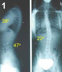
BEFORE TREATMENT STARTING POINT X-ray prior to the brace (15 Jan 1998). Patient’s birth date: 9 Jun 1983. Risser 2. First x-ray with the brace, shoelift R=+0.5cm. Starting date: 17 Feb 1998. Age: 14 yr 7 mth. Risser 2. WEANING POINT FOLLOW-UP POINT X-ray taken at 1 yr 3 mth (27 May 1999) into brace treatment. Age: 15 yr 11 …
Case Study 3
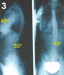
BEFORE TREATMENT STARTING POINT X-ray prior to the brace (7 Oct 1997). Patient’s birth date: 1 Apr 1983. Risser 3. First x-ray with the brace. Starting date: 7 Oct 1997. Age: 14 yr 6 mth. Risser 3. WEANING POINT FOLLOW-UP POINT X-ray taken at 1 yr 8 mth (3 Jun 1999) into brace treatment. Age: 16 yr 2 mth Risser …
Case Study 4
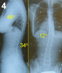
BEFORE TREATMENT STARTING POINT X-ray prior to the brace (15 Jul 1997). Patient’s birth date: 2 Mar 1982. Risser 2. First x-ray with the brace. Starting date: 6 Aug 1997. Age: 15 yr 5 mth. Risser 2. WEANING POINT FOLLOW-UP POINT X-ray taken at 2 years (29 Jul 1999) into brace treatment. Age: 17 yr 5 mth Risser3. X-ray taken …